
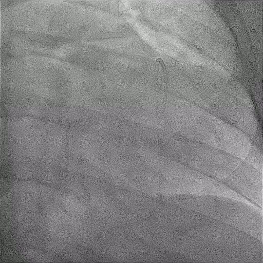
What is pericardiocentesis?
The heart sits inside a thin, tough sac called the pericardium. Normally there is only a teaspoon or two of fluid inside it, but illness can cause a large amount to collect. Because the sac cannot stretch quickly, the fluid presses on the heart and stops it from filling properly, which makes you breathless, dizzy and weak; when it is severe this is called tamponade and it is an emergency. Pericardiocentesis is the procedure in which that fluid is drained with a needle and a fine tube passed through the chest wall, using ultrasound to guide it safely.
Why is it done?
The first reason is to relieve pressure on the heart, which often produces immediate improvement in breathing and blood pressure. The second is to find out why the fluid collected: it is sent to the laboratory to test for infection, including tuberculosis, for cancer cells and for signs of inflammation, and those results guide your further treatment. Small collections that cause no symptoms are usually watched rather than drained.
What happens on the day?
The procedure may be planned or done urgently at the bedside. You lie propped up, a drip is placed and your heart rhythm and blood pressure are monitored. Ultrasound is used to find the safest window to the fluid, usually just below the breastbone or on the left side of the chest. The skin and deeper tissues are numbed thoroughly with local anaesthetic, and light sedation may be given. A needle is advanced under ultrasound guidance until fluid appears, then a soft drain is placed over a wire and the needle removed. The fluid is drained slowly and the drain is stitched in place and connected to a bag. It usually takes twenty to forty minutes, and you may feel pressure or a brief sharp sensation but should not have significant pain.
What to expect afterwards
Most people feel better very quickly. The drain is usually left in for a day or two so that any fluid that reforms can escape, and it is removed once the drainage has almost stopped and the ultrasound looks satisfactory. You will be monitored in a high care or ward bed, and repeat ultrasound scans check that the fluid has not returned. The site is tender for a few days and may bruise. Treatment then focuses on the underlying cause, which may involve antibiotics, tuberculosis treatment, anti-inflammatory medication or oncology care. If the fluid keeps coming back, a surgical drainage procedure or a pericardial window may be recommended.
Possible complications
When ultrasound guidance is used, serious complications are uncommon, but they can be significant. The needle can injure the heart muscle or a coronary artery, causing bleeding that occasionally needs surgery, or puncture the lung, causing air to leak around it and requiring a chest drain. Other possible problems are disturbance of the heart rhythm, injury to the liver or an abdominal organ, infection introduced along the drain, and the fluid reaccumulating after removal of the drain. In very ill patients the procedure carries a risk to life, but leaving significant tamponade untreated is far more dangerous. Your doctor will explain the balance in your case, often quickly if the situation is urgent.
This information does not replace a consultation. Please discuss your own risks and options with your cardiologist.